Case Description :
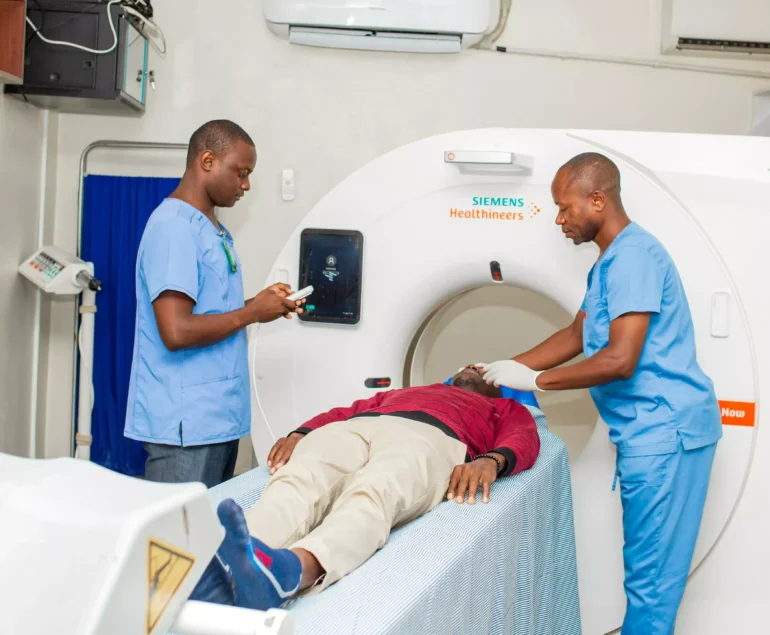
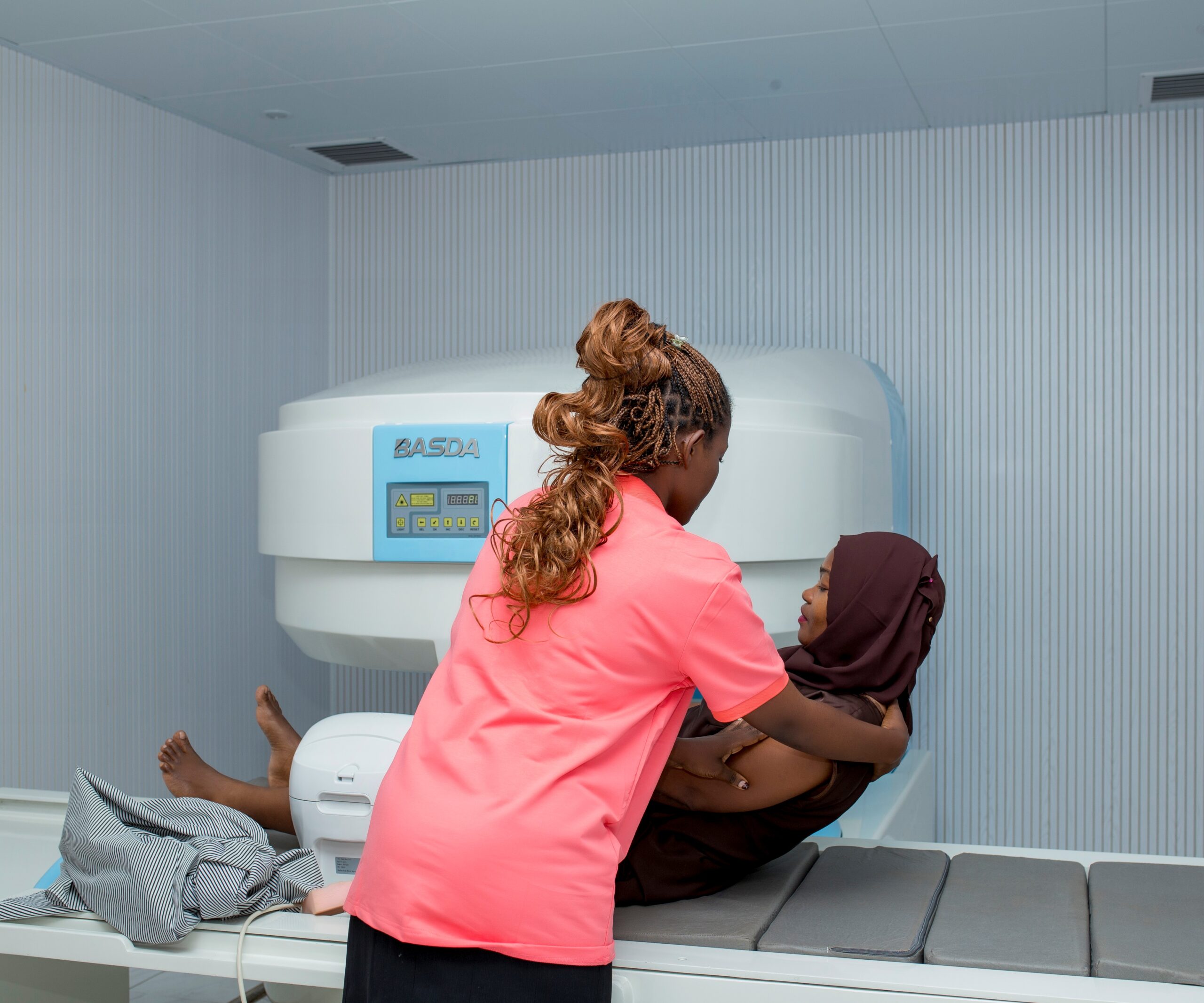
When a patient undergoes an MRI (Magnetic Resonance Imaging) scan, the process begins with the radiographer positioning the patient and configuring the MRI machine according to the clinical request. The machine generates detailed cross-sectional images of the body using magnetic fields and radio waves. (Acquisition of Images, Interpretation and Analysis, Reporting)
What We Do
- Orthopedics (for musculoskeletal injuries and joint problems)
- Oncology (for tumor detection, staging, and follow-up).
- Cardiology (for cardiac MRI and vascular imaging)
- Gastroenterology (for abdominal MRI such as liver or pancreas imaging)
- Urology / Gynecology (for pelvic scans).

Medical Laboratory and Specialist Services Supporting MRI
- Hematology (blood counts to check for anemia, infection)
- Biochemistry (renal and liver function tests before contrast use).
- Tumor markers (for oncology cases).
- Microbiology (if infection is suspected).
- Neurology / Neurosurgery (for brain and spinal conditions)
Final Results :

A well-structured MRI report provides clear discrete final results rather than vague descriptions. For example:
Clinical Indication: Chronic headaches, rule out space-occupying lesion.
Findings:
No intracranial mass lesion identified.
Ventricular system is normal in size and configuration.
No abnormal enhancement after contrast.
Impression / Final Result: Normal MRI Brain. No evidence of tumor, hemorrhage, or demyelination.