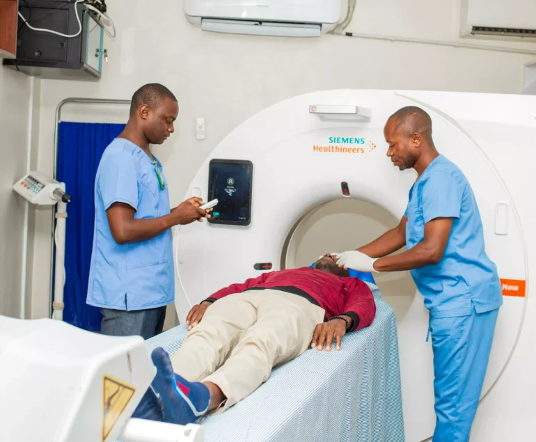
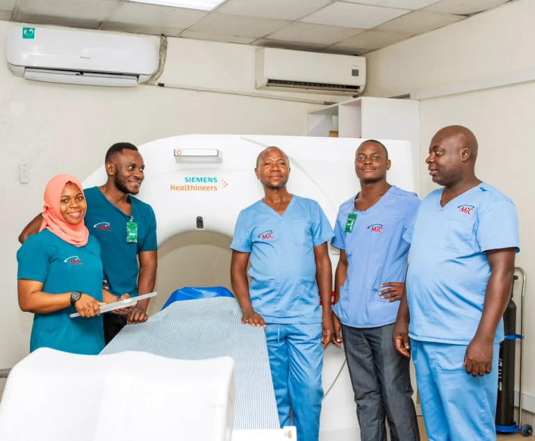
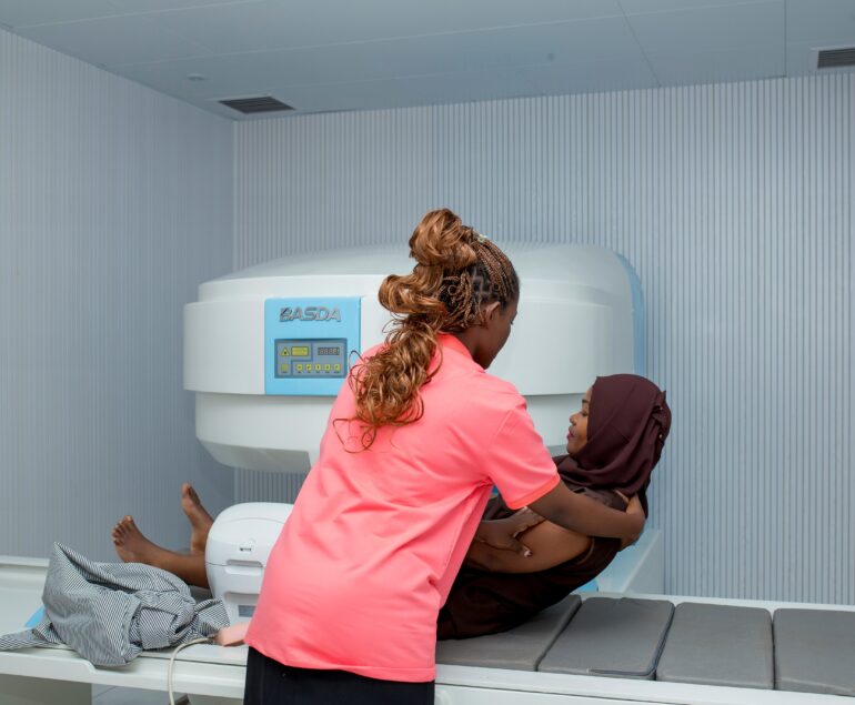
Case Description :
Ultrasound (sonography) is a safe, non-invasive imaging technique that uses high-frequency sound waves to produce real-time images of internal organs, soft tissues, and blood vessels. Medical imaging consultants provide this service across a wide range of diagnostic and interventional cases, supporting physicians in clinical decision-making.
Common Cases Where Ultrasound is Used
- Abdominal Imaging – liver, gallbladder, pancreas, spleen, kidneys, urinary bladder.
- Obstetric & Gynecological – pregnancy assessment, fetal growth and wellbeing, uterus, and ovarian evaluation.
- Cardiac (Echocardiography) – heart function, valve assessment, and chamber evaluation.
- Vascular Studies – Doppler ultrasound to assess blood flow in arteries and veins (e.g., detecting clots, blockages, aneurysms).
- Musculoskeletal Imaging – tendons, ligaments, muscles, and joints for tears, inflammation, or fluid collections.
- Small Parts & Superficial Structures – thyroid, breast, testes, and lymph nodes.
- Interventional Guidance – guiding needle biopsies, fluid drainage, or catheter placements.
What We Do :

Step-by-Step Breakdown of the Procedure
- Patient Preparation
- Positioning
- Application of Gel
- Scanning
- Post-Examination Care
Results & Reporting
Normal Findings: Clear visualization of organ structure, normal size, shape, and vascularity.
Abnormal Findings:
- Masses, cysts, or tumors.
- Inflammation, infection, or fluid collections.
- Blockages, narrowing, or abnormal blood flow in vessels.
- Fetal abnormalities or growth restriction in obstetric cases.
Report Delivery: A radiologist/consultant interprets the images and provides a detailed written report to the referring physician.
Outcome: The results guide diagnosis, monitor disease progression, or assist in planning interventions.


